Integrating R&D, production, sales and technical servicesModern high-tech enterprises
Tel400-876-2378
Tel400-876-2378

Hot tags:ADA adenosine deaminase test kit (peroxidase method) Cys-C cystatin C test kit (latex enhanced immunoturbidimetry) LDL-C Low Density Lipoprotein Cholesterol Test Kit (direct method surfactant clearance method) PCT procalcitonin test kit (latex enhanced immune turbidimetry) Bf Factor B Test Kit (Latex Enhanced Immunoturbidimetry)
PHONE
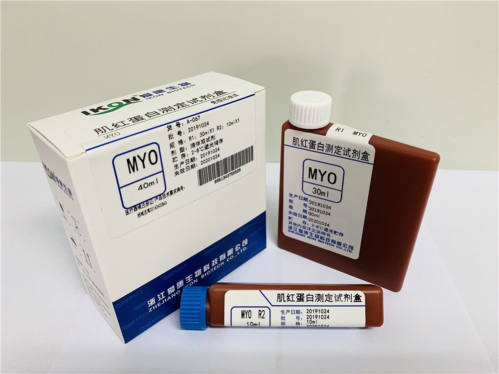
400-876-2378Myoglobin (Mb) is a protein that stores and distributes oxygen in mammalian cells (mainly muscle cells). It is composed of a polypeptide chain and a coenzyme heme, with a relative molecular weight of 16700 and containing 153 amino acid residues. The deproteinized myoglobin, excluding heme, is called globulin. It shares significant homology with the subunits of hemoglobin (a-globin chain and P-globin chain) in the amino acid sequence, and their conformation and function are also extremely similar.
Myoglobin is a binding protein composed of a peptide chain and a heme subunit. It is a protein that stores oxygen in muscles, and its oxygen saturation curve is hyperbolic. Myoglobin is present in muscles and is particularly abundant in the myocardium. The tertiary structure of sperm whale myoglobin was elucidated by Kendrew using X-ray diffraction in 1960, making it the world's first described protein tertiary structure. Due to the direct correlation between the tertiary structure and the biological function of proteins, and the high difficulty of analyzing the tertiary structure, this work has received high praise from the academic community. Myoglobin has 8 segments α- Spiral Zone Each α- The helical region contains 7-24 amino acid residues, called A, B, C... G, and H peptide segments, respectively. Myoglobin has 1-8 helical regions, with a non helical region (also known as the helical region) at the corner of the peptide chain, including a non helical region with 2 amino acid residues at the N end and 5 amino acid residues at the C end. Polar amino acids such as Pro, Ile, Ser, Thr, Asn located at the inflection point are distributed on the molecular surface, and there is a pocket shaped hole inside, in which heme resides. Heme is an iron porphyrin compound, which is composed of four pyrroles connected by four methoxynyl groups to form a large ring, with Fe2+residing in the ring. Connection of iron with porphyrin rings and peptide chain amino acid residues: The two propionic acid side chains on iron porphyrin are connected to the positive charges on the two basic amino acid side chains in the peptide chain in the form of ionic bonds. The Fe2+of heme forms coordination bonds with the nitrogen atoms of four pyrrole rings, with one of the other two coordinating bonds binding to F8 histidine and one binding to O2. Therefore, heme maintains a stable position in this hole.
This conformation is very beneficial for oxygen transport and storage functions, while also maintaining stability of heme in the peptide chain. However, excessive exercise, fatigue, solar radiation, air pollution, smoking, pesticides, etc. can produce excessive free radicals. Free radicals, also known as "free radicals" in chemistry, are atomic groups containing an unpaired electron. Due to the fact that electrons in chemical bonds must appear in pairs when atoms form molecules, free radicals seize an electron from other substances everywhere to form stable substances. In chemistry, this phenomenon is called "oxidation". Reactive oxygen species in the body have certain functions, such as immune and signal transduction processes. But excessive reactive oxygen species can lead to destructive behavior, leading to damage to normal cells and tissues in the human body, while myoglobin is an oxygen-rich chain protein that is more susceptible to free radical attacks. Being attacked by free radicals can lead to various diseases, such as heart disease, Alzheimer's disease, Parkinson's disease, and tumors, which are closely related to the oxidation of myoglobin. In addition, more reactive oxygen species cause nucleic acid mutations, which are the root cause of human aging and disease. The clinical significance of myoglobin determination. Serum myoglobin can be used as the earliest and most sensitive indicator for the diagnosis of acute myocardial infarction (AMI). But its specificity is poor, and diseases such as skeletal muscle injury, trauma, and renal failure can all lead to its elevation. Although positive Myo cannot diagnose AMI, it can be used as an important indicator for early exclusion of AMI diagnosis. If negative Myo, myocardial infarction is basically excluded, and it can also be used for the diagnosis of reinfarction. In combination with clinical practice, if Myo rises again, it should be considered as reinfarction or infarction extension. Elevated: seen in early acute myocardial infarction, acute muscle injury, muscular dystrophy, muscle atrophy, polymyositis, acute or chronic renal failure, severe congestive heart failure, and long-term shock. It can increase 1.5 hours after myocardial infarction, but returns to normal within 1-2 days. 1. Elevated blood levels: hypothyroidism, hyperaldosteronism, renal insufficiency, malignant hyperthermia, and severe exercise. 2. Elevated levels in urine: porphyria, hemoglobinuria, hematuria, etc. 3. Elevated myoglobin in blood and urine: seen in acute myocardial infarction, angina pectoris, cardiogenic shock, cardiomyopathy, muscle diseases (progressive muscular dystrophy, polymyositis, myasthenia gravis), etc.
Copyright©en.zjikon.com
( copy )Zhejiang Aikang Biotechnology Co., Ltd
Powered by 
hot city:
 CN
CN