Integrating R&D, production, sales and technical servicesModern high-tech enterprises
Tel400-876-2378
Tel400-876-2378

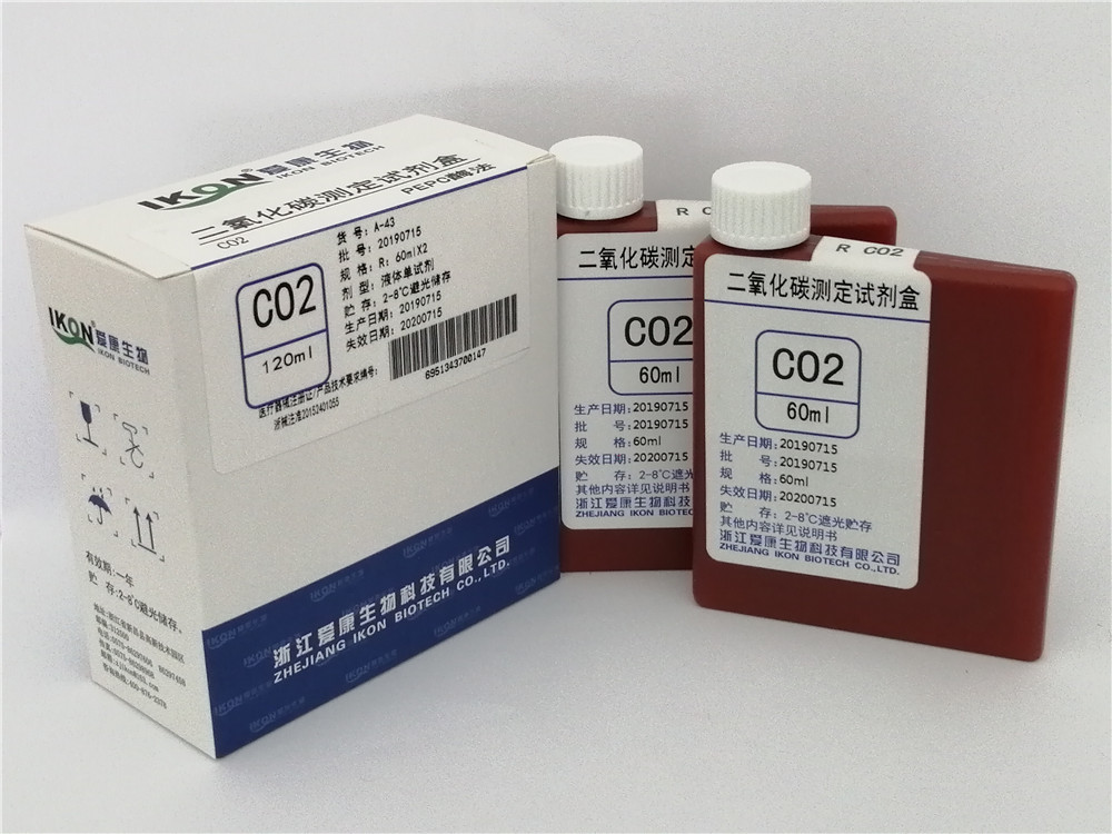
Hot tags:ADA adenosine deaminase test kit (peroxidase method) Cys-C cystatin C test kit (latex enhanced immunoturbidimetry) LDL-C Low Density Lipoprotein Cholesterol Test Kit (direct method surfactant clearance method) PCT procalcitonin test kit (latex enhanced immune turbidimetry) Bf Factor B Test Kit (Latex Enhanced Immunoturbidimetry)
PHONE
400-876-2378The B-factor assay kit is mainly based on the principle of enzyme-linked immunosorbent assay (ELISA), which detects the content of B-factor in the sample through antigen antibody specific binding reaction. Its core working principle and process are as follows:.
Enzyme labeling technology amplifies signals
Enzyme labeled secondary antibodies (such as horseradish peroxidase, HRP) are added to form a "solid-phase antibody B factor enzyme labeled secondary antibody" complex with already bound factor B.
. Enzyme markers can catalyze substrate coloration, and the concentration of factor B can be quantitatively analyzed by color intensity.Color reaction and quantitative analysis
Adding a substrate (such as TMB), the enzyme catalyzes the substrate to produce a colored product (such as blue), which turns yellow under acidic conditions.
. The color depth is directly proportional to the concentration of factor B. The absorbance (OD value) is measured by an enzyme-linked immunosorbent assay (ELISA) reader, and a standard curve is drawn to calculate the content of factor B in the sample.2. Operating Procedure (Taking Double Antibody Sandwich Method as an Example)
Sample Preparation
Serum/Plasma: Centrifuge the supernatant to avoid repeated freezing and thawing.
.Tissue homogenate: Rinse the tissue with pre cooled PBS, cut and homogenize, centrifuge to obtain the supernatant.
. Cell culture supernatant: Centrifuge to remove cell debris and take the supernatant for detection.Sample addition and incubation
Add standard samples (gradient dilution) and test samples to the coated plate, and incubate to allow the antigen to bind to the solid-phase antibody.
. Wash to remove unbound components and reduce non-specific interference.Add enzyme labeled secondary antibody
Add HRP labeled secondary antibody and incubate to form a complex.
. Wash again to remove unbound enzyme-linked antibodies.Color development and termination
Add substrates A and B, and incubate in the dark for color development.
. Add termination solution (such as sulfuric acid), and the color changes from blue to yellow.Result determination
OD value was measured at 450nm wavelength using an enzyme-linked immunosorbent assay (ELISA) reader.
. Calculate the concentration of factor B in the sample based on the standard curve.III. Key Technical Features
High Sensitivity
Enzyme catalyzed signal amplification, capable of detecting factor B as low as ng/mL, suitable for early disease diagnosis or micro sample analysis.
.High specificity
Antigen antibody specific binding, avoiding cross reactivity and ensuring accuracy of results.
.Easy to operate
Standardized process, matching kit containing all necessary components, suitable for rapid detection in clinical or scientific research.
.Quantitative analysis
Absolute quantification is achieved through standard curves, providing objective data support for disease diagnosis.
.IV. Application Scenarios
Complement system function evaluation: detecting the level of factor B reflects the activity of the bypass pathway, assisting in the diagnosis of hereditary angioedema (HAE), paroxysmal nocturnal hemoglobinuria (PNH) and other diseases.
. Autoimmune disease monitoring: Overactivation of factor B in patients with systemic lupus erythematosus (SLE) may exacerbate inflammation, and detecting its levels can help evaluate disease activity. Drug development: Verify the efficacy of complement system inhibitors (such as Eculizumab) and monitor changes in factor B before and after treatment.Copyright©en.zjikon.com
( copy )Zhejiang Aikang Biotechnology Co., Ltd
Powered by 
hot city:
 CN
CN