Integrating R&D, production, sales and technical servicesModern high-tech enterprises
Tel400-876-2378
Tel400-876-2378

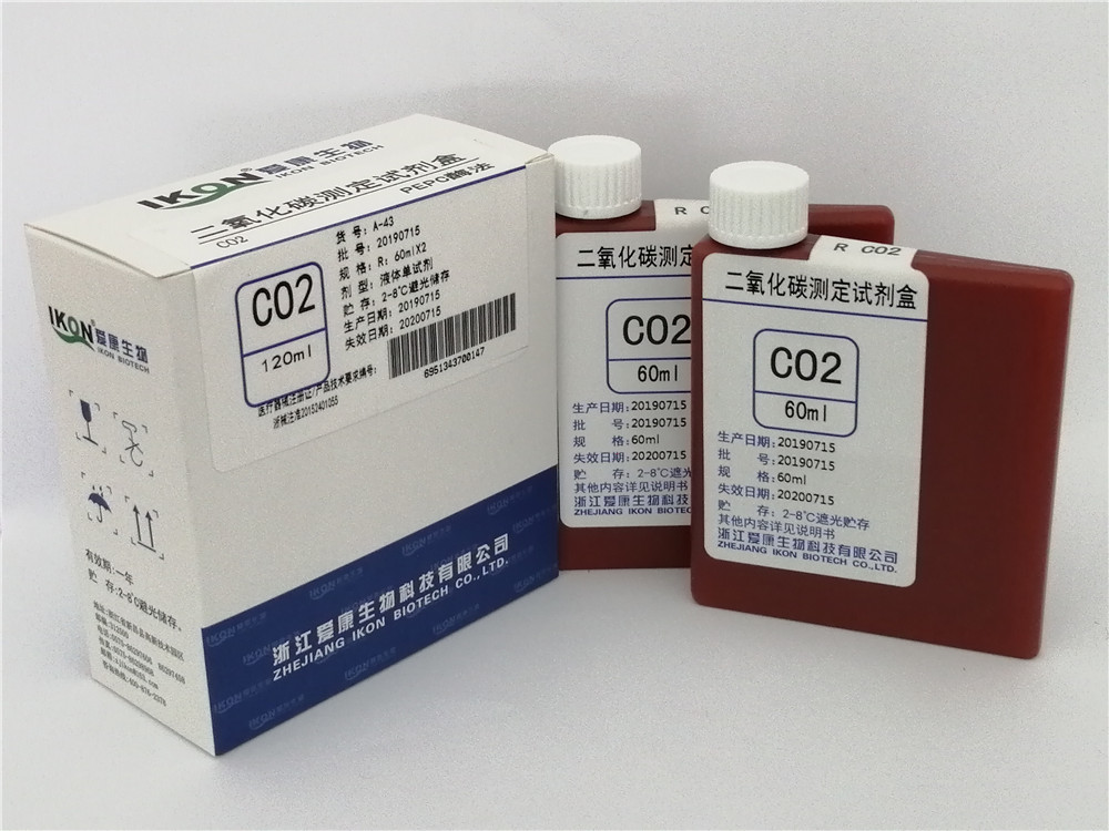
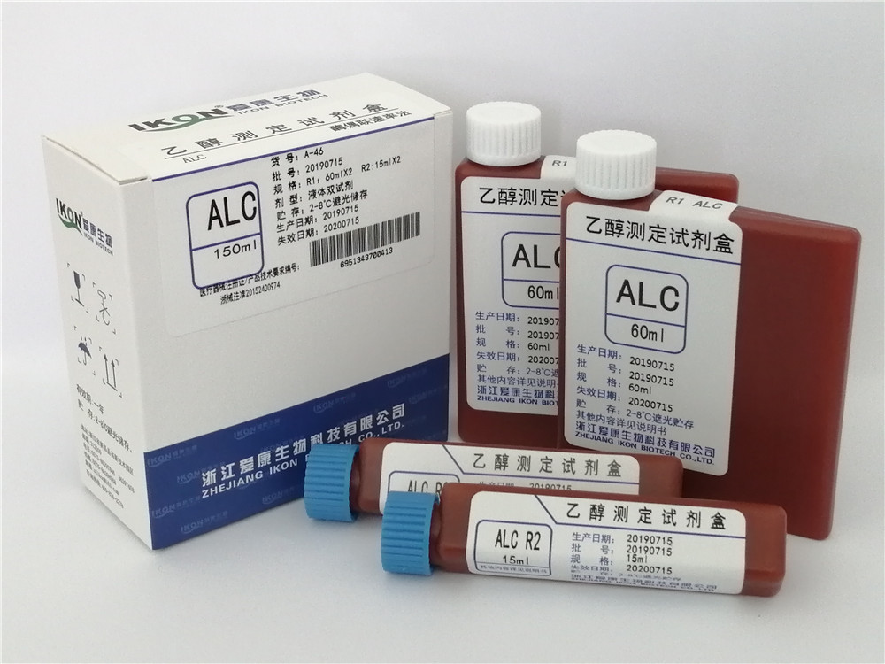
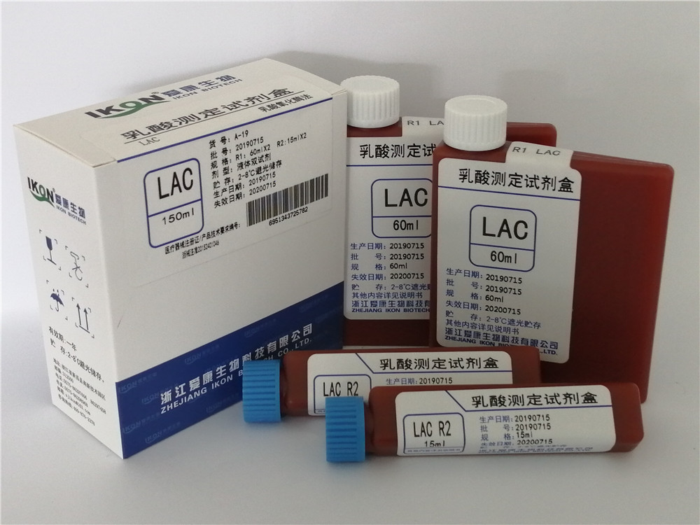
Hot tags:ADA adenosine deaminase test kit (peroxidase method) Cys-C cystatin C test kit (latex enhanced immunoturbidimetry) LDL-C Low Density Lipoprotein Cholesterol Test Kit (direct method surfactant clearance method) PCT procalcitonin test kit (latex enhanced immune turbidimetry) Bf Factor B Test Kit (Latex Enhanced Immunoturbidimetry)
PHONE
400-876-2378The usage method of anti trypsin assay kit varies depending on the type of kit (such as ELISA kit, activity detection kit) and the test object (human, mouse, rat samples), but the core steps usually include sample processing, reagent preparation, sample addition, incubation, washing, color development and termination, result determination and calculation. The following are general operating steps and precautions:
1. Sample processing
Serum/plasma:
Collect blood using test tubes that do not contain pyrogens and endotoxins. Serum sample: After being left at room temperature for 2 hours or overnight at 4 ℃, centrifuge at 1000 × g for 20 minutes and collect the supernatant.
Plasma samples: Use EDTA or heparin as anticoagulants, centrifuge at 2-8 ℃ and 1000 × g for 15 minutes within 30 minutes after collection, and collect the supernatant.
.Avoid hemolysis and hyperlipidemia. If there are particles in the serum, centrifuge or filter.
.Tissue homogenate:
Rinse the tissue with pre cooled PBS (0.01M, pH=7.4) to remove residual blood.
. After weighing, cut the tissue into pieces and add PBS (protease inhibitor can be added) at a ratio of 1:9 (weight to volume). Grind thoroughly on ice, or perform ultrasonic fragmentation and repeated freeze-thaw cycles to lyse cells. Centrifuge at 5000 × g for 5-10 minutes and collect the supernatant for detection.Cell supernatant:
Centrifuge at 1000 × g for 20 minutes and collect the supernatant.
. If detecting intracellular components, dilute the cell suspension with PBS to around 1 million/ml, repeat freeze-thaw cycles, and centrifuge to obtain the supernatant.Storage:
If not used immediately, the sample should be packaged and stored at -70 ℃ to avoid repeated freezing and thawing.
. Thawing should be carried out at room temperature to ensure that the sample is evenly and fully thawed.II. Reagent Preparation
Kit Composition:
Includes enzyme-linked immunosorbent assay (ELISA) plate, standard, sample diluent, detection antibody HRP, washing buffer, substrate A, substrate B, stop buffer, etc.
. The components of different reagent kits may vary and need to be prepared according to the instructions.Reagent preparation:
Wash buffer: Dilute with distilled water according to the instructions (such as 1:20 or 1:50).
.Standard: Dilute the series according to the instructions, use and prepare immediately, do not store.
.Other reagents, such as biotinylated antibodies, affinity chain enzyme HRP, etc., are usually ready to use.
.3. Sample addition
Set standard and sample wells:
Add 50 μ L of standard at different concentrations to each standard well.
. Add 50 μ L of the test sample to the sample well; Blank holes are not added.Precautions for sample addition:
When adding samples, try not to touch the hole wall and gently shake and mix well.
. If the sample concentration is too high, it needs to be diluted with sample diluent before adding the sample, and the calculation should be multiplied by the corresponding dilution factor.IV. Incubation
Incubation conditions:
In addition to blank wells, 100 μ L of horseradish peroxidase (HRP) labeled detection antibody is added to each well of the standard and sample wells.
. Seal the reaction well with a sealing film and incubate at 37 ℃ in a water bath or constant temperature box for 60 minutes (the specific time should be adjusted according to the instructions of the reagent kit).Incubation precautions:
Ensure that all reagents reach room temperature (20-25 ℃) before use.
. During the incubation process, do not let the micropores dry out.Fifth, Washing
Washing steps:
Discard the liquid and pat dry on absorbent paper.
. Fill each well with washing solution (350 μ L), let it stand for 1 minute, shake off the washing solution, and pat dry on absorbent paper. Repeat washing the board 5 times (or use a board washing machine to wash the board).Washing precautions:
Insufficient washing can lead to accuracy errors and incorrect increase in OD values.
.Make sure to absorb as much liquid as possible from the well before adding the substrate.
VI. Color Development and Termination
Color Development:
Add 50 μ L of substrate A and B to each well, and incubate at 37 ℃ in the dark for 15 minutes (the specific time should be adjusted according to the instructions of the kit).
. The substrate color solution should be colorless or very light in color, and the substrate solution that has turned blue cannot be used.Termination:
Add 50 μ L of termination solution to each well, and measure the OD value of each well at a wavelength of 450nm within 15 minutes (specific time adjusted according to the kit instructions).
. The order of adding the termination solution should be as consistent as possible with the order of adding the substrate solution.7. Determination and Calculation of Results
Determination of OD Value:
Measure the absorbance (OD value) of each well in sequence using an enzyme-linked immunosorbent assay (ELISA) reader at a wavelength of 450nm.
. The measurement should be carried out immediately after adding the termination solution.Calculate sample concentration:
Use the OD value of the measured standard as the horizontal axis and the concentration value of the standard as the vertical axis. Draw a standard curve on a coordinate paper or with relevant software, and obtain a linear regression equation.
. Substitute the OD value of the sample into the equation and calculate the concentration of the sample. If the sample has been diluted, it needs to be multiplied by the corresponding dilution factor to obtain the actual concentration.Tags:Antitrypsin,assay,kit,
Copyright©en.zjikon.com
( copy )Zhejiang Aikang Biotechnology Co., Ltd
Powered by 
hot city:
 CN
CN