Integrating R&D, production, sales and technical servicesModern high-tech enterprises
Tel400-876-2378
Tel400-876-2378

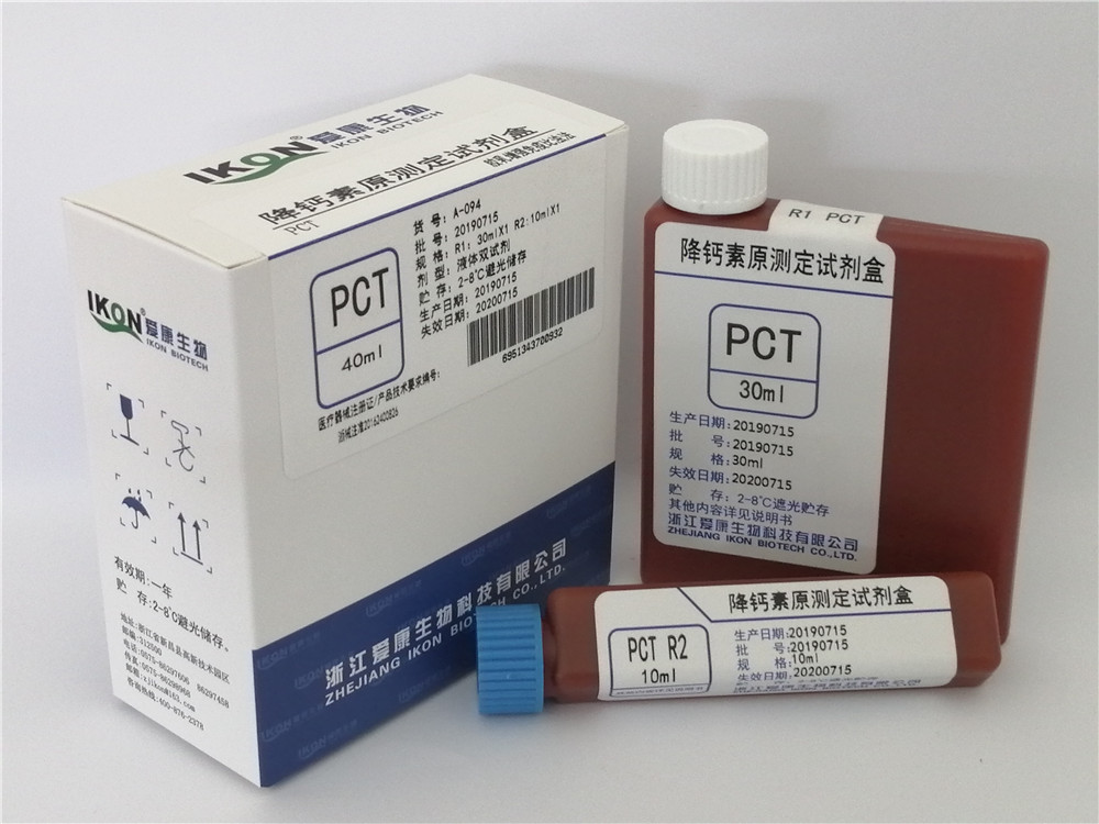
Hot tags:ADA adenosine deaminase test kit (peroxidase method) Cys-C cystatin C test kit (latex enhanced immunoturbidimetry) LDL-C Low Density Lipoprotein Cholesterol Test Kit (direct method surfactant clearance method) PCT procalcitonin test kit (latex enhanced immune turbidimetry) Bf Factor B Test Kit (Latex Enhanced Immunoturbidimetry)
PHONE
400-876-2378At present, there are many laboratory methods to detect PCT, which can be qualitative and quantitative. The procalcitonin test usually has the following methods:
1. Radioimmunoassay
The synthetic polyclonal antibody was used to specifically recognize and link the amino acid procalcitonin. This method can detect the serum PCT of normal people with a reliable sensitivity of 4pm/mL. It can detect the mixture of free PCT, binding PCT and calcitonin gene-related peptide precursor, but can not distinguish the above three substances. The detection time of this method is long (19~22h), and the use of radioactive elements is limited.
2. Double antibody sandwich immunochemiluminescence assay (ILMA)
Using double monoclonal antibodies, one of which is calcitonin antibody and the other is anti-calcitonin antibody, respectively bind to the calcitonin and anti-calcitonin sites of PCT molecules, which can eliminate cross reactions. One antibody is marked by the cursor, and the other unmarked antibody is fixed on the inner wall of the test tube. During the reaction, the two antibodies combine with PCT molecules to form a sandwich complex, and the light emitting part is located on the surface of the reaction tube. The method is simple, specific and sensitive. The lower limit of the determination is 0.1 ng/mL, and the results can be obtained within 2 hours.
3. Colloidal gold colorimetry (B.R.A.H.M.SPCT-Q-semi-quantitative rapid test)
Using colloidal gold technology, including colloidal gold labeled anti-calcitonin monoclonal antibody and anti-calcitonin polyclonal antibody used as coating, when the sample (serum or plasma) is added into the sample hole, the gold-labeled monoclonal antibody combines with the PCT in the sample to form the gold-labeled antigen-antibody complex. The complex moves on the reaction membrane and combines with the anti-calcitonin antibody fixed on the membrane to form a larger complex. When the concentration of PCT exceeds 0.5ng/mL, the complex displays red, and the depth of red is proportional to the concentration of PCT, and the concentration range of PCT can be obtained by comparing with the standard color palette. The results were divided into four levels: normal<0.5ng/mL; Slightly increased>0.5ng/mL; Significantly higher than 2ng/mL; Significantly higher than 10ng/mL. The method is fast, simple and easy to observe.
4. Transmission immune turbidimetry
The PCT in the sample reacts with the PCT monoclonal antibody in the reagent in an antigen-antibody manner, which increases the turbidity of the reaction solution. Within a certain range, the turbidity of the reaction solution is linear with the amount of added human antigen. The absorbance value of the reaction solution can be measured at the wavelength of 6O0nm using a biochemical analyzer or other optical detection instrument. The absorbance value of the reaction solution is in direct proportion to the measured PCT concentration. The method is simple, rapid, automatic and suitable for batch detection. In 2005, PCT immunoturbidimetric kits developed in China were supplied, which provided convenient conditions for the wide application of PCT. Although the methodology and clinical application of immune transmission turbidimetry need further verification, its application prospect is very promising.
This is the end of today's article. I want to share it with you here. I hope you can enjoy it and pay more attention to us. We will continue to provide better articles.

Copyright©en.zjikon.com
( copy )Zhejiang Aikang Biotechnology Co., Ltd
Powered by 
hot city:
 CN
CN